


// Workers AI · dad joke modeWhat did labial glands say? We lip service.
| Labial glands | |
|---|---|
 The labial coronary arteries, the glands of the lips, and the nerves of the right side seen from the posterior surface after removal | |
| Details | |
| Artery | Inferior labial |
| Identifiers | |
| Latin | glandulae labiales oris |
| TA98 | A05.1.02.014 |
| TA2 | 2813 |
| FMA | 71613 |
| Anatomical terminology | |
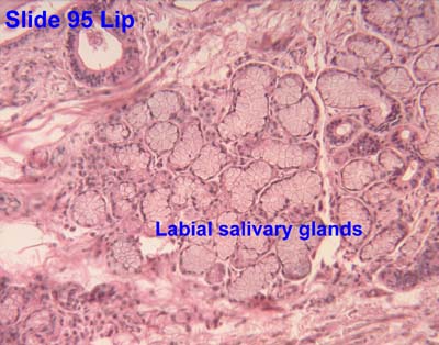
The labial glands are minor salivary glands situated between the mucous membrane and the orbicularis oris around the orifice of the mouth.
They are circular in form, and about the size of small peas; their ducts open by minute orifices upon the mucous membrane.
Like the parotid and buccal glands, the labial glands are innervated by parasympathetic fibres that arise in the inferior salivatory nucleus, travel with the glossopharyngeal nerve and lesser petrosal nerve to the otic ganglion, where they synapse and then continue to the labial glands. Sympathetic innervation is mediated by postganglionary fibres which arise in the superior cervical ganglion and pass through the otic ganglion without synapsing.
References
[edit] This article incorporates text in the public domain from page 1111 of the 20th edition of Gray's Anatomy (1918)
External links
[edit]- Histology image: 95_08 at the University of Oklahoma Health Sciences Center


 | This anatomy article is a stub. You can help Wikipedia by adding missing information. |